Scientists from Sun Yat-sen University’s School of Biomedical Engineering have made a significant breakthrough in the field of bone regeneration.
By developing advanced tubular scaffolds using electrospun membranes, they have created a cutting-edge solution to promote the healing of critical skull defects.
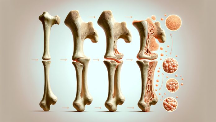
These scaffolds, designed to mimic the structure of natural bone, provide an ideal environment for stem cells to flourish and accelerate the healing process, marking a major step forward in tissue engineering and regenerative medicine.
Addressing critical bone defects
Critical-sized bone defects have long been a major challenge in the medical world. Traditional treatments, such as autografts and allografts, often face limitations, including the scarcity of donors, size mismatches, and potential immune rejection.
These issues have hindered the widespread use of these methods for bone repair. However, the growing field of bone tissue engineering offers a promising solution.
Adipose-derived stem cells (ADSCs), which are easily accessible and possess strong osteogenic (bone-forming) capabilities, have attracted significant attention for their potential in bone regeneration.
While injecting ADSCs directly into defect sites often results in a short survival time, combining them with scaffold materials has proven to enhance retention and improve bone regeneration.
Researchers are now exploring new ways to develop scaffolds that mimic the natural structure of bone, utilising methods like electrospinning and 3D printing.
Innovative tubular scaffolds for bone regeneration
The team at Sun Yat-sen University tackled these challenges head-on by developing multilayer composite nanofibrous membranes made from polycaprolactone (PCL), poly(lactic-co-glycolic acid) (PLGA), and nano-hydroxyapatite (HAp).
These materials, created using electrospinning technology, are engineered to replicate the structure of bone. When shaped into tubular scaffolds, they create an optimal environment for adipose-derived stem cells (rADSCs) to promote bone regeneration.
The scaffolds not only simulate bone structure but also enhance the proliferation and osteogenic differentiation of rADSCs, meaning they help these stem cells transform into bone-forming cells more effectively.
In laboratory and animal studies, the scaffolds demonstrated remarkable results in promoting bone growth and healing.
With a bilayer thickness ratio of 1:2 and an initial total thickness of 2.5 μm, these materials can spontaneously transform into 3D scaffolds when exposed to certain conditions, adding to their practicality in medical applications.
The future of bone regeneration
The success of these scaffolds points to a bright future for bone regeneration treatments. The research has shed light on the mechanisms behind how these scaffolds, combined with growth factors like VEGF and BMP-2, promote bone formation.
By integrating both chemical signals and physical properties, these advanced scaffolds have the potential to revolutionise bone defect repair.
Further research is needed to optimise the design of these fibrous scaffolds and explore the mechanisms by which mesenchymal stem cells (MSCs) promote bone regeneration.
However, the results thus far are highly promising, offering a new approach to treating bone defects that could soon be applied in clinical settings.


