The Department of Clinical Neuroscience at Karolinska Institutet in Sweden has developed a new imaging method to examine the dopamine system in the brain of patients with Parkinson’s disease.
Dopamine is a neurotransmitter responsible for the control of movements. The cells that produce dopamine are located in an area of the brainstem, the substantia nigra. Through nigro-striatal projections, dopamine is then secreted from the axonal terminals into the striatum, the area of the brain that plays an important function in regulating motor activity. In Parkinson’s disease, dopamine cells degenerate and their loss is responsible for the motor symptoms that characterise the disorder, such as bradykinesia, rigidity and tremor.
How DAT imaging with high-resolution PET can be used as a mapping tool
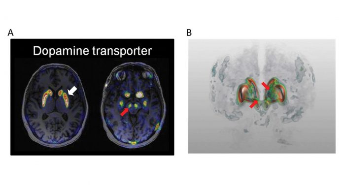
Using a brain imaging technique known as Positron Emission Tomography (PET), we have measured the availability of the dopamine transporter protein (DAT) that regulates the dopamine levels in the brain. Since 2008 the high-resolution research tomograph (HRRT) has been operational at the PET-Centre of the Department of Clinical Neuroscience at Karolinska Institutet. The HRRT is the PET systems with highest resolution (~2mm) for imaging of the brain. High-resolution PET combined with the best-in-class DAT imaging agent, [18F]FE-PE2I, enables the imaging of the DAT in the dopamine cell bodies, on the axons and on the nerve terminals (Fig. 1). DAT imaging with high-resolution PET can be used as a biomarker to map the entire dopamine system in vivo.
A PET study has been conducted at Karolinska Institutet on 20 patients with early Parkinson’s disease and 20 age- and gender-matched control subjects. In Parkinson’s patients the DAT availability in the nerve terminals was found to be 70% lower as compared with control subjects, whereas the DAT levels remained relatively more preserved (30% lower in patients versus controls) in cell bodies and nerve fibres.
These results suggest that in the early stages of the disease there is a prevalent loss of striatal terminals whereas the majority of the dopamine cells are still viable and their function could be restored with correct treatment. The method we have developed can in future assist in the early diagnosis of Parkinson’s disease and in the follow-up of the disease. DAT imaging can also be used as a biomarker in clinical trials of new medicines and treatment strategies. Future studies will examine patients with more advanced disease, in order to examine the relationship between DAT and clinical variables such as motor symptoms and the various stages of the disease.
References
Varrone A, Sjoholm N, Eriksson L, Gulyas B, Halldin C, Farde L. Advancement in PET quantification using 3D-OP-OSEM point spread function reconstruction with the HRRT. Eur J Nucl Med Mol Imaging. 2009;36:1639-1650.
Fazio P, Svenningsson P, Cselényi Z, Halldin C, Farde L, Varrone A. Nigrostriatal dopamine transporter availability in early Parkinson’s disease. Mov Disord. 2018;33(4):592-599.
Andrea Varrone
Associate Professor
Department of Clinical Neuroscience
Karolinska Institutet
+46 8 517 750 43
andrea.varrone@ki.se


